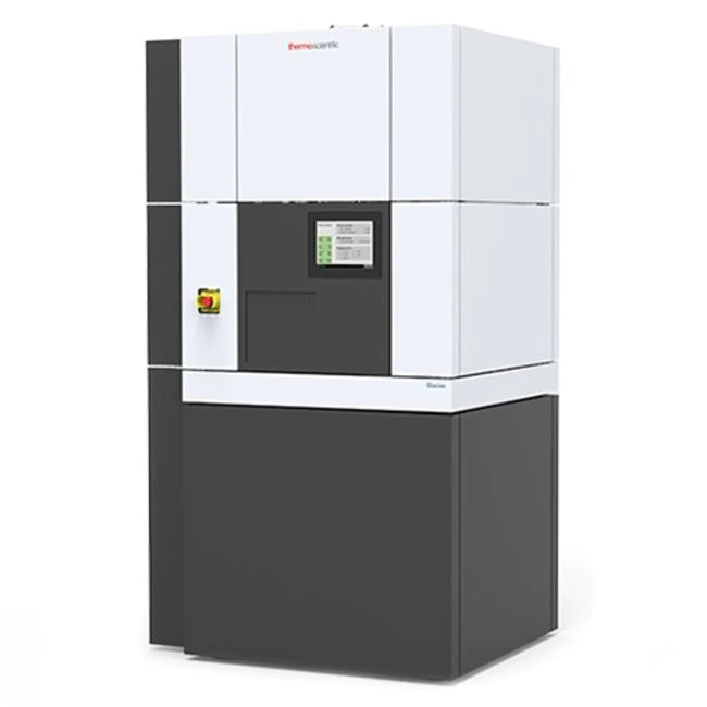
Researchers at IU School of Medicine are about to welcome their very own cryo-electron microscope (cryo-EM) to the school. Scientists are already very familiar with the cutting-edge cryo-EM technology, but the onslaught of the COVID-19 pandemic actually made some members of the public appreciate the many benefits of this Noble Prize-winning technology too.
As COVID-19 began to emerge in the middle of January 2020, a complete DNA sequence of the disease it causes, known as severe acute respiratory syndrome coronavirus 2 (SARS-CoV-2) was published. Only a month later, using cryo-EM, the structure of the spike protein associated with SARS-CoV-2 was determined and published by scientists at the University of Texas in Austin and the National Institutes of Health.
“Knowledge of the structure makeup has been pivotal in identifying treatments that bind to the protein in order to find cures and treatments against the virus that has claimed so many U.S. lives, said Yuro Takagi, PhD, Associate Professor of Biochemistry and Molecular Biology. “Determining the structure of the spike protein at this unprecedented speed was only possible thanks to cryo-EM technology.”
Now this revolutionary technology is coming to IU School of Medicine. “Hopefully we never find ourselves in another pandemic situation. But if we do, we will have a convenient cryo-EM right here on campus to get us in the fight for a cure,” said Takagi.
The ability to purchase the cryo-EM for the school called the Glacios™ came as a result of a high-end instrumentation grant, or more simply known as an “S10 grant” from the NIH designed to help NIH-funded investigators to obtain cutting-edge tools that advance NIH research. Takagi was awarded with the S10 grant and is the technical director and technical advisory committee member for the IU School of Medicine's new cryo-EM facility.
“Yuro was instrumental in bringing cryo-EM to the school,” said Quyen Hoang, PhD, Associate Professor of Biochemistry and Molecular Biology. "None of this would have happened without him."
A cryo-EM on campus is a very big deal in the school’s own drug discovery efforts, as it will allow school researchers to map out how disease proteins of interest and potential drug candidates interact with each other at near atomic resolution, thereby helping school researchers to design better compounds.
“We know that LRRK2 is a protein, which when mutated causes familial Parkinson's disease. Because it is a large protein, it is very suitable for cryo-EM, but because its shape is widely variable, we were never able to get a uniform sample required to obtain the high-resolution data with a cryo-EM. Using our own cryo-EM will allow us to screen for various conditions in order to get a suitable sample of LRRK2 and obtain the needed high-resolution data,” said Hoang.
IU School of Medicine researchers are at the forefront of Parkinson’s disease research; once they have a compound or antibody to treat the disease, they will also use the cryo-EM to gain insight as to how it will bind to the LRRK2 disease target.
Cryo-EMs also overcome the limitations of earlier technologies such as X-ray crystallography which requires a researcher to first grow crystals of the protein they are studying. Nuclear Magnetic Resonance (NMR) technology is limited to work with smaller protein molecules as opposed to larger proteins associated with structural biology. Cryo-EM freezes a protein as it exists in its natural state and then allows a scientist to see it at molecular and atomic structural levels.
With exceptional capabilities also come a big price tag. The cryo-EM costs nearly $3 million to own and operate. Funding for the machine has been provided by the NIH S10 grant, the school, and the Lilly Endowment, which is subsidizing some of the costs of the cryo-EM through its INCITE collaboration with the school.
“We are going to price use of the cryo-EM with competitive rates to ensure everyone has access to use it,” said Takagi. “We want Lilly researchers to have access, as well as any other scientist across the state who can benefit from the capabilities of our machine. We also plan to set up all the necessary computational infrastructure to analyze data from the cryo-EM to accommodate the needs of school users and beyond.”
The Glacios™ is expected to arrive in mid-May and be located on the first floor of the medical sciences building. It will form the basis of a new IUSM Center for Electron Microscopy core, which will be led by Hoang. The use of the Glacios™ is complementary to the Titian Krios™ Cryo-EM technology that school researchers were also instrumental in bringing to the state back in 2019. The Titian Krios™ is housed at Purdue University.
For more information about the cryo-EM facilities, please contact Yuro Takagi at ytakagi@iu.edu or Quyen Hoang at qqhoang@iu.edu.
