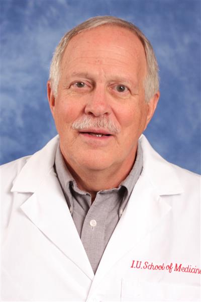
George E. Sandusky, DVM, PhD
Research Professor of Pathology & Laboratory Medicine
Professor, Pathology and Laboratory Medicine, Indiana University School of Medicine
Professor, Pathology, Purdue University
Director of the IU Simon Cancer Center Tissue Bank
Associate Director of the Indiana CTSI Specimen Storage Facility (SSF)
Co-Director, INBrain (Indiana Center for Biomarker Research in Neuropsychiatry)
Bio
Dr. George Sandusky Professor, Dept Pathology and Laboratory Medicine, IU School of Medicine finished DVM at The Ohio State University in 1971, completed pathology residency and received his doctorate in pathology at Louisiana State University in 1981. Dr. Sandusky is recognized for his leadership and technical contributions that have impacted programs targeting cancer and cardiovascular disease in the Indiana University Department of Pathology and Laboratory Medicine has broad knowledge in anatomic pathology and in tissue banking procedures and quality assurance focused on collection, processing, storage, and qc of cancer specimens and the normal adjacent tissue since the IU tissue bank inception in 1996. Through the years he has worked on all types of issues relating to informed consent, collection, storage, tissue requests, quality control issues, and database issues.
Dr. Sandusky serves as the Director and PI for the IU Simon Cancer Center Tissue bank which has a very large, well annotated, high quality tissue bank with very high quality samples collected (Sandusky et al 2006, Sandusky et al, 2009, Tarvin and Sandusky 2014).
Dr. Sandusky has mentored and taught approximately 125 undergraduate, graduate and medical students over the past 25 years in several different fall, spring, summer and yearlong internship programs requiring a completed poster and / or oral presentation. There have been approximately 25 poster presentations at the AACR during the past 10 years.
Dr. Sandusky’s lab has capabilities to analyze whole slide digital imaging for clinical and research cases as a service for both whole slide digital images as well as tissue microarrays. Historically, pathologists have hand read H&E or immunohistochemically-stained slides. With the advent of technology, we are now capable of using analysis software to quanitfy positive pixel readings to validate the hand score by a pathologist.
Additionally, the lab offers pathology reviews for the cases and utilizes virtual microscopy to view slides which allows the ability to pan, zoom, and explore the slide via a computer system.
The lab focuses on Aperio Whole Slide Digital Imaging and quantitative image analysis and has the capabilities to use Image Pro Plus for single field image analysis, Aperio positive pixel algorithms, Indica Halo algorithms, and quPath as well as Orbit Whole Slide Image Analysis. The lab has around five to eight undergraduate and graduate research assistants.
Key Publications
| Year | Degree | Institution |
|---|---|---|
| 1980 | PhD | Louisiana State University |
| 1978 | MS | The Ohio State University |
| 1971 | DVM | The Ohio State University |
| 1967 | BS | Ohio University |
Immunohistochemistry, Molecular Pathology, Cancer Research, Digital Whole Slide Image Analysis
Desc: Ohio State Distinguished Alumni Award
Scope:
Date: 2016-05-01
