
28-year-old with low platelet count

Hematology

Authors: Agnes Davenport FNP-C and Nichole LePage, MD, Indiana University School of Medicine

Summary of clinical history

The patient is a 28-year-old female with a history of systemic lupus erythematosus (SLE) complicated by nephritis and severe cutaneous manifestations managed by multiple immunosuppressant medications. She was transferred to the emergency department after outpatient rheumatology labs revealed hemoglobin of 5.9 GM/dL [12-15] and platelet count of 10 k/mm3 [150-450].

Patient had a known history of iron deficiency anemia, menorrhagia and pancytopenia. She was admitted by the hematology service to undergo evaluation for severe anemia and thrombocytopenia. Her clinical examination was unremarkable.

Measurement Patient Reference Range / Expected Result
Hemoglobin 5.9 GM/dL 12.0-15.0
Platelet 10 k/mm3 150-450
MCV 95fL 81-99
Reticulocyte 5.2% 0.5-2.5
Haptoglobin <6 mg/dL 30-200
Serum ferritin 157.9 ng/mL 10-106
Serum Iron 64mCg/dL 50-212
Total Iron-Binding Capacity 260mCg/dL 250-400
PT 12.2 seconds 9.3-13.5
INR 1.03 0.73-1.17
LDH 773 U/L 140-271
Creatinine 1.02 mg/dL 0.6-1.2
Folate >23 ng/mL >5.9
Vitamin B12 151pg/mL 200-1000
DAT Negative Negative


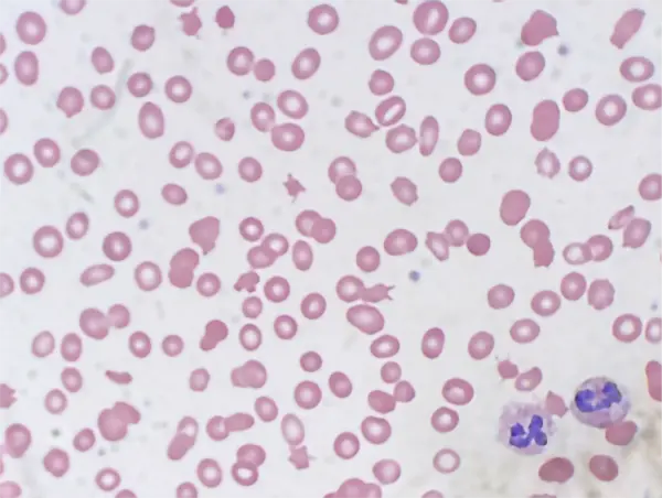
Microscopic findings
A peripheral blood smear was consistent with laboratory findings of anemia and thrombocytopenia. Abnormalities of red blood cell morphology were appreciated including dacrocytes, macrocytes, microcytes, and ovalocytes. There were many schistocytes appreciated on the sample collected on the day of admission. Schistocytes in combination with an elevated lactate dehydrogenase are consistent with microangiopathic hemolytic anemia.

Case of the month 6, photo of cells


Clinical Course
Patient was started on corticosteroids due to concerns of immune-related pancytopenia etiologies and folinic acid for possible methotrexate toxicity. She was noted to have vaginal bleeding consistent with her known history of menorrhagia and this could have contributed to her anemia in addition to hemolysis. Tests for anticardiolipin and anti-beta-2-glycoprotein antibodies were below clinically significant thresholds, her serum methotrexate level resulted at <0.03 mCmol/L, and her ADAMTS13 activity resulted at <5%.

Neurological and physical exam remained within normal limits. Her platelet count remained below 20 k/mm3 despite two platelet transfusions. Her anemia responded appropriately to red blood cell transfusions despite persistent heavy vaginal bleeding.

Test yourself: What diagnosis would you give this patient?

Incorrect diagnosis

Incorrect diagnosis

Incorrect diagnosis

Incorrect diagnosis

Diagnosis by authors

Discussion of case

Thrombotic thrombocytopenic purpura (TTP), a thrombotic microangiopathy, is characterized by severe thrombocytopenia and microangiopathic hemolytic anemia. This is caused by a dysfunction of ADAMTS13, a metalloprotease enzyme, either by immune-mediated antibody binding and destruction or by an inherited mutation. In healthy hemodynamics, ADAMTS13 cleaves von Willebrand factor (vWF) multimers. When the enzyme is removed from circulation by immune destruction or dysfunctional, vWF multimers excessively bind and aggregate platelets, leading to thrombotic complications and decreased circulating platelets.

Microvascular thrombi lead to organ ischemia most commonly in the kidneys and brain. Thrombi in smaller vasculature causes the schistocytes seen on peripheral smear as narrowed lumens cause mechanical stress and shearing on the red blood cell membrane. The laboratory values that support hemolysis include undetectable haptoglobin, elevated reticulocyte count, and elevated lactate dehydrogenase (LDH). The classic presentation for TTP is a pentad of symptoms: fever, thrombocytopenia, hemolytic anemia, renal injury, and neurological symptoms. Due to effective and prompt initiation of therapy very few patients with diagnosed TTP develop all five symptoms.

Due to the risk of thrombi and subsequent organ ischemia rapid initiation of plasma exchange with donor plasma replacement is considered an emergent procedure for the reduction of morbidity and mortality in presentations concerning for TTP prior to laboratory confirmation. This treatment both removes antibodies to ADAMTS13 and provides functional ADAMTS13 enzyme in patients with immune-mediated TTP. A study in 1991 revealed a decrease in mortality from >90% to 10-20% when plasma exchange treatment was started in immune-mediated cases of TTP.

This patient had multiple potential diagnoses and underlying bleeding that complicated her clinical picture. Her underlying SLE is the most likely cause for her secondary acute immune TTP. Her ADAMTS13 activity resulting below 5% is well below the expected 60% at the low end of normal activity. A reflex test for ADAMTS13 antibody resulted at 40 U/mL [<11 u/ml]. She received two weeks of daily plasma exchange with donor plasma replacement followed by rituximab infusion. In addition to daily plasma exchange therapy, immunosuppressive medications are part of TTP treatment to reduce production of ADAMTS13.

Key differential diagnosis

Iron deficiency Anemia
Despite her severe anemia and history of iron deficiency anemia (IDA), her elevated reticulocyte count on presentation is not supportive of IDA in which the reticulocyte count is typically low due to insufficient iron stores for erythropoiesis. Other findings that dispute IDA include a normal mean corpuscular volume (MCV), thrombocytopenia, elevated ferritin and normal total iron binding capacity. Further, a diagnosis of IDA does not explain the hemolysis seen in this case.

B12 Vitamin Deficiency
Although her vitamin B12 is below the reference range the evidence that supports it has not affected erythropoiesis include the normal MCV, elevated reticulocyte count, and normal nuclear segmentation of the neutrophils seen on peripheral smear. B12 deficiency further does not explain the hemolysis seen in this case.

Drug induced (Methotrexate) bone marrow suppression
An adverse event with immunosuppressive medication such as methotrexate is often associated with polypharmacy or underlying liver and kidney disease. While this patient was prescribed and taking methotrexate, her serum drug levels were below detectable levels and her measured kidney function was within normal limits. Bone marrow suppression could concurrently cause anemia and thrombocytopenia, the hemolysis seen in the patient would remain unexplained.

Catastrophic Antiphospholipid Syndrome
Catastrophic antiphospholipid syndrome (CAPS) is another form of thrombotic microangiopathy that may present similarly to TTP. Additionally CAPS is associated with SLE and results in thrombocytopenia and hemolysis. The differentiation between CAPS and TTP in this patient’s case is best supported by the lack of clinically significant antiphospholipid antibodies including anticardiolipin and anti-beta-2-glycoprotein and the support of TTP via decreased ADAMTS13 activity. 

References

  1. Connelly-Smith L, Alquist CR, Aqui NA, Hofmann JC, Klingel R, Onwuemene OA, et al. Guidelines on the use of therapeutic apheresis in clinical practice—evidence-based approach from the Writing Committee of the American Society for Apheresis: the Ninth Special Issue. J Clin Apher. 2023;38(2):77-278. doi:10.1002/jca.22043

  2. Sukumar S, Lämmle B, Cataland SR. Thrombotic thrombocytopenic purpura: pathophysiology, diagnosis, and management. J Clin Med. 2021;10(3):536. doi:10.3390/jcm10030536

  3. Scully M, Carter MA, Subhan MO. Thrombotic thrombocytopenic purpura: celebrating 25 years of ADAMTS13. Blood. Published online March 2, 2026. doi:10.1182/blood.2025030791

  4. Rock GA, Shumak KH, Buskard NA, Blanchette VS, Kelton JG, Nair RC, Spasoff RA. Comparison of plasma exchange with plasma infusion in the treatment of thrombotic thrombocytopenic purpura. Canadian Apheresis Study Group. N. Engl. J. Med. 1991;325: 393-397.